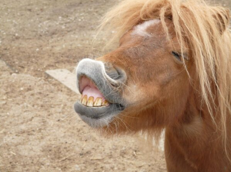
A Lifetime of Teeth — How Horse Teeth & Care Changes with Age
A horse’s teeth tell its life story. The experienced dentist reads in them not just age, but diet, past illness, management history, and — sometimes — years of unrecognised pain.
A horse’s dental story begins before birth and continues until the animal’s final years. Unlike humans, horses are hypsodont — their teeth are exceptionally tall relative to their crown, with a long reserve of tooth buried below the gum line. Over a lifetime, this reserve erupts gradually to compensate for the constant grinding of fibrous forage. Understanding this process helps explain why equine teeth health are so closely tied to age.
Birth to six months — the milk teeth. Foals are born with, or rapidly develop, a set of 24 deciduous (milk) teeth: incisors and premolars only. These first teeth are smaller, whiter, and less robust than permanent teeth. They establish the foal’s first bite and begin the chewing patterns that will shape jaw development throughout life.
Six months to five years — the great replacement. This is the most turbulent dental period of a horse’s life. All 24 milk teeth are gradually shed and replaced by permanent counterparts, and 12 entirely new permanent cheek teeth (the molars, which have no deciduous predecessors) erupt at the back of each arcade. The shedding process follows a predictable sequence:
Eruption timeline of permanent teeth
- 2–2.5 years — First permanent incisors (centrals) erupt; first permanent premolars push out their caps
- 2.5–3 years — First molars complete eruption; wolf teeth appear (typically upper jaw, around 18 months to 3 years)
- 3–3.5 years — Second permanent incisors (intermediates) and second premolars erupt
- 3.5–4 years — Second molars in full wear
- 4–4.5 years — Third incisors (corners) and third premolars erupt; canine teeth emerge in males
- 4.5–5 years — Third (rear) molars erupt; by five, the permanent arcade is essentially complete
During this transition, dental caps — the remnants of shed milk teeth sitting atop erupting permanent teeth — are a frequent complication for equine teeth health. If a cap fails to dislodge naturally, it traps the permanent tooth beneath and can cause significant discomfort and misalignment.
Five to fifteen years — the working prime. Once the permanent dentition is established, the focus shifts from eruption to wear management. A horse in this age bracket typically has a full, well-aligned arcade with plenty of reserve tooth remaining. Annual dentistry is usually sufficient, with floating correcting the inevitable sharp points that form through normal chewing. This is the period when problems are most preventable and least expensive to manage.
Fifteen to twenty-five years — the gradual reduction. As the reserve crown is used up, the tooth becomes shorter and the roots shallower. The protective infundibulae (the dark central pits visible on the chewing surface) wear away, and the dental star — a yellow-brown mark of secondary dentine — becomes increasingly visible. Teeth that have served for decades begin to loosen slightly in their sockets, and the risk of periodontal disease rises. Many horses in this bracket benefit from twice-yearly examinations.
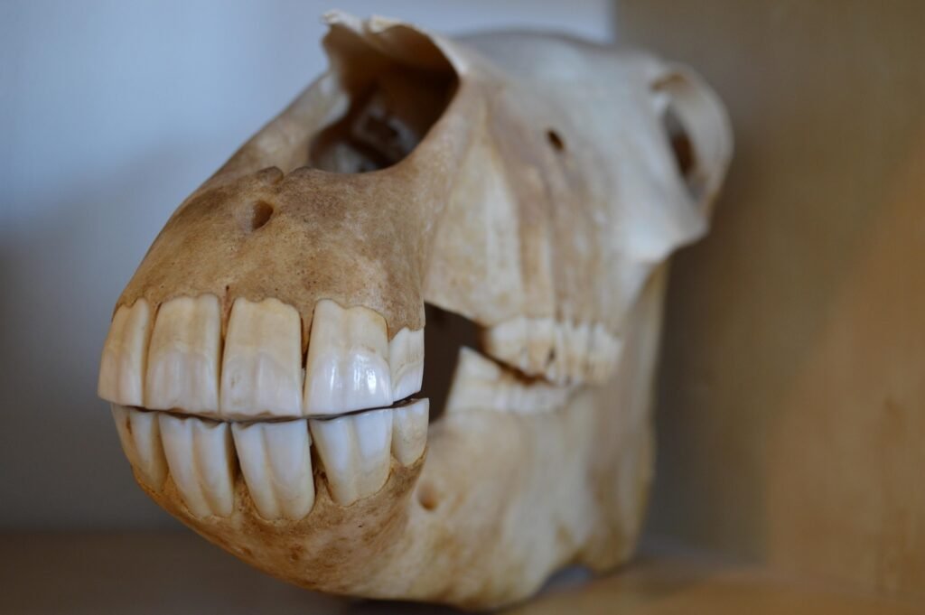
What does the Equine Dentist Actually Do?
An equine dental examination is far more involved than a quick peek inside the mouth. A qualified equine dental technician or vet — often called an equine dentist — follows a systematic process that typically takes 30 to 60 minutes and requires specialist equipment.
Sedation first. Most horses are given a mild sedative, commonly detomidine or romifidine, before the exam. This is not merely for convenience — a fully conscious horse cannot safely hold its mouth open long enough for a thorough assessment, and stress can mask or mimic clinical signs.
The speculum. A Hausmann’s gag (or mouth speculum) is fitted between the upper and lower incisors to hold the mouth open. Without it, the cheek teeth — the large molars and premolars at the back — are inaccessible.
Visual and tactile inspection. Using a bright headlamp and a dental mirror, the practitioner inspects all 36 to 44 teeth in a mature horse — incisors, canines (in males), wolf teeth, premolars, and molars. They use gloved hands to feel along each tooth for sharp edges, loose teeth, abnormal angles, and soft-tissue swelling of the gums and cheeks.
Percussion and probing. Tapping individual teeth can reveal pain responses that indicate infection or fracture. Periodontal probes measure the depth of gaps between tooth and gum, identifying pockets where feed material can pack and ferment.
Assessment of wear and occlusion. The practitioner evaluates how the upper and lower arcades meet — called the occlusal surface. Horses’ teeth erupt continuously throughout their lives and wear against each other; any disruption to that process creates the problems described in the next section.
Age estimation. The teeth are also a biological clock. The degree of wear on the incisors, the shape of the tables (biting surfaces), and the appearance of specific marks — Galvayne’s groove, the dental star — allow an experienced dentist to estimate a horse’s age to within a year or two.
Common problems with horse teeth
Horse teeth are not static objects — they erupt, shift, wear unevenly, and respond to diet, age, and genetics. Below are the conditions an equine dentist encounters most frequently.
Sharp Enamel Points
The most common finding. The upper cheek teeth overhang the lower arcade, creating razor-sharp edges on the outer (buccal) surface. These lacerate the cheek and tongue, causing pain particularly when a bit is in use.
Wave Mouth
The occlusal surface undulates in a wave pattern — some teeth overgrown, others worn flat. Causes inefficient chewing and forces the jaw into abnormal positions, cascading into neck and back tension.
Step Mouth
A single tooth is dramatically taller than its neighbours, often because the opposing tooth is missing or damaged. Feed cannot be ground properly, and the overgrown tooth can injure the opposing gum.
Hooks and Ramps
Hooks form on the front edge of the first upper cheek tooth or the rear edge of the last lower cheek tooth when the jaw sits slightly forward or back. They restrict jaw movement and can cause bit resistance and head-tilting.
Wolf Teeth
Small vestigial teeth that erupt just in front of the premolars, usually in the upper jaw. They sit exactly where a bit rests and frequently cause bit pain, evasion, and head-shaking in ridden horses.
Periodontal Disease
Bacterial infection of the gums around individual teeth. Feed packs into widened gum pockets, ferments, and destroys the supporting structures. Can lead to tooth loss and, in severe cases, sinusitis.
Diastema (Gaps)
Abnormal spaces between adjacent teeth that trap feed material. Often painful, and can be a primary driver of quidding (dropping partially chewed food) and weight loss in older horses.
Fractures & Caries
Teeth can fracture from trauma or feed-related decay (equine caries). Fractured teeth expose sensitive pulp canals, cause severe pain, and are a route for infection into the tooth root and adjacent sinuses.
Stomatitis
Stomatitis simply means inflammation of the mucous membranes that line the mouth — the gums, inner cheeks, tongue, and soft palate. It is not a single disease but a clinical sign that can arise from a variety of underlying causes READ MORE…
Periodontal Disease
Bacterial infection of the gums around individual teeth. Feed packs into widened gum pockets, ferments, and destroys the supporting structures. Can lead to tooth loss and, in severe cases, sinusitis.
Older horses (typically 15+) are more likely to present with loosening teeth, severe periodontal disease, and missing teeth that disrupt the whole arcade. Very young horses (2–5 years) go through a complex shedding of 24 deciduous (“milk”) teeth, and retained caps or abnormal eruption patterns are common during this period.
Warning Signs for dental issues in horses
Horses are stoic animals that rarely telegraph pain in obvious ways. The signs of dental discomfort and poor equine teeth health are often interpreted as behavioural, training, or digestive problems. Knowing what to look for — and what to do about it — can make an enormous difference to a horse’s welfare.
Warning Signs in Your Horse
- Quidding — dropping balls of half-chewed hay or grass from the mouth while eating
- Weight loss despite adequate forage, because the horse cannot properly process food
- Slow eating, eating only on one side of the mouth, or avoiding hard feed
- Facial swelling, particularly below the eye or along the jaw — may indicate an abscess or sinusitis
- Foul smell from the mouth or nostrils — often a sign of infection or decaying trapped feed
- Undigested grain in droppings — the horse is swallowing feed before adequate chewing
- Bit resistance, head-tossing, or tilting in a ridden horse with no previous history of it
- Head-shaking or reluctance to take a contact, especially on one rein
- Nasal discharge from one nostril — a classic sign of a tooth root abscess draining into a sinus
What can you proactively do?
- Book a dental examination at least once a year for adult horses, and every six months for horses under five or over fifteen. Many problems are far easier — and cheaper — to treat when caught early.
- Ensure ad-lib or regular access to good-quality long-stem forage. Constant chewing of fibrous material creates the lateral jaw movement that wears teeth evenly. Horses on very soft or short-cut diets are more prone to dental irregularities.
- Watch your horse eat. Five minutes of observation at feed time will often reveal quidding, one-sided chewing, or slow eating before it becomes clinically obvious.
- Never ignore ridden resistance. If a horse that previously worked well suddenly resists the bit, shakes its head, or fights one rein, rule out dental pain before assuming a training problem. And, PLEASE, check the bit you are using for the correct fit!
- Maintain records. Keep a dental diary noting the date of each examination, findings, and treatment. This is invaluable for tracking progression in older horses and is increasingly expected by insurance companies.
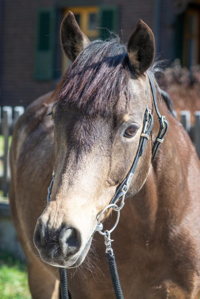
How are problems with equine teeth health treated?
Treatment ranges from routine maintenance that takes a few minutes to surgical extraction requiring referral to an equine hospital. The vast majority of dental work is performed in the field, with the horse sedated and the practitioner working by headlamp and feel.
Floating (Rasping) The cornerstone of equine dentistry. Manual or motorised rasps — called floats — are used to file down sharp enamel points and reduce overgrowths. Motorised equipment (power floats) allows faster, more precise work on difficult-to-reach cheek teeth. The goal is to produce a smooth, level occlusal surface on which upper and lower teeth meet evenly.
Wolf Tooth Removal Wolf teeth are extracted routinely, usually in young horses before bitting begins. The tooth and its root are loosened with an elevator — a fine, flat instrument — and lifted cleanly from the socket. The procedure takes minutes and the socket heals within a fortnight. Most horses are ridden normally within a few days.
Retained Cap Removal When a milk tooth fails to shed naturally as the permanent tooth pushes through, the practitioner loosens and removes the retained cap. Left in place, caps cause discomfort, misdirect the erupting tooth, and can lead to the malocclusions described above.
Periodontal Flushing Pockets infected by periodontal disease are flushed with antiseptic solution to remove packed food material and bacteria. In mild cases this is sufficient; more advanced disease may require the pocket to be widened to allow ongoing drainage and easier cleaning by the owner with a syringe at home.
Diastema Management Wide diastemata are widened further — counterintuitively — using burrs, so that feed falls through rather than packing in. Narrow gaps may be widened or treated with periodic flushing. Severe cases sometimes benefit from dental impression material placed in the gap to prevent food accumulation between visits.
Tooth Extraction When a tooth is irreparably fractured, severely infected, or causing lasting damage to surrounding structures, extraction is necessary. Simple, mobile teeth can be removed in the field under sedation and nerve block. Firmly rooted cheek teeth require more involved techniques: a dental pick and molar cutters in experienced hands, or referral for repulsion (pushing the tooth up through a hole drilled in the sinus) or minimally invasive transbuccal extraction at a hospital.
Sinus Treatment Tooth root infection frequently spreads into the adjacent paranasal sinus, causing sinusitis with one-sided nasal discharge. If antibiotics alone fail, the sinus may need to be trephined (a small hole drilled into the skull under local anaesthetic) and flushed, often alongside extraction of the offending tooth.
Post-Extraction Care After extraction, the empty socket is flushed and packed with dental putty or gauze to exclude feed. Owners are given a syringe to flush the socket with dilute chlorhexidine solution twice daily for several weeks. Once healed, the opposing tooth (which no longer has anything to grind against) must be rasped at every subsequent visit to prevent it from overgrowing.